World's Smallest 3D Camera Developed For Endoscopic Brain Surgery

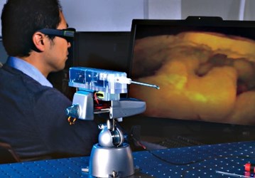
NASA scientists are working with researchers at the Skull Base Institute (SBI) in Los Angeles to develop a “super instrument,” an endoscope with a 3D camera that will allow neurosurgeons to better navigate brain tissue during tumor resections. The Multi Angle Rear Viewing Endoscopic Tool (MARVEL) is designed to assist and further perfect minimally invasive surgery for skull base tumors.
In 2010, Gary Gallia, neurosurgeon at Johns Hopkins Medical Center, said in a podcast that 30-35 percent of brain tumors are skull base tumors, centrally located and extremely difficult to access through traditional craniotomies. Neuro research of the past decade has focused on perfecting neuroendoscopic procedures that are less invasive, offering patients safer surgeries and quicker recovery times.
In that interview, Gallia commented that further development of the technique would focus on imaging technology, particularly 3D technology that would allow surgeons to see around the tumor and locate it in relation to surrounding sensitive and fragile tissue, such as the carotid artery or optic nerve.
Now, a team of scientists at SBI, led by Hrayr Shahinian, is working with NASA’s Jet Propulsion Laboratory (JPL) in Pasadena to develop MARVEL. Shahinian and his team aim to produce the most state-of-the-art tools for endoscopic brain surgery, a goal which requires them to provide the best possible 3D image resolution with the smallest possible instrument.
According to SBI scientists, the best current neuroendoscopic tools can only offer a panoramic view, 30-70 degrees from a fixed angle, displayed as a 2D image via an attached monitor. In comparison, MARVEL offers a 120-degree view and a 3D image of the tumor that gives the surgeon depth perception and better visibility.
Furthermore, typical stereo imaging endoscopes have dual cameras. In order to miniaturize their technology even further, SBI and JPL scientists reduced the number of camera lenses from two to one. To achieve the 3D effect, the camera has two apertures with different-colored filters. When the images are sent to the monitor through a radio transmitter fitted to the end of the scope, they’re merged to create a 3D image, scientists said in a JPL press release.
The finished laboratory prototype camera is 4 mm in diameter and 15 mm long, the smallest camera ever developed for this type of application. The highly bendable neck of the scope makes MARVEL extremely maneuverable in tight spaces.
The team’s next step will be to develop a clinical prototype to submit to the FDA for approval. Scientists say the design will undergo further refinement to ensure that it is both effective and feasible in real-world medical settings.
“As a skull base surgeon with a specific vision of endoscopic brain surgery,” said Shahinian, “it has been a privilege and a great personal honor working with the JPL team over the past eight years to realize this project.”
