Unwanted Side Effect Becomes Advantage In Photoacoustic Imaging

By Beth Miller
Biomedical engineer Lihong Wang, PhD, and researchers in his lab work with lasers used in photoacoustic imaging for early-cancer detection and a close look at biological tissue. But sometimes there are limitations to what they can do, and as engineers, they work to find a way around those limitations.
Wang, the Gene K. Beare Distinguished Professor of Biomedical Engineering in the School of Engineering & Applied Science at Washington University in St. Louis, and Junjie Yao, PhD, a postdoctoral research associate in Wang’s lab, found a unique and novel way to use an otherwise unwanted side effect of the lasers they use — the photo bleaching effect — to their advantage.
The results were published online Jan. 10 in Physical Review Letters.
The researchers use an optical microscopy method called photoacoustic microscopy to take an intensely close look at tissues. The laser beam is a mere 200 nanometers wide. However, the center of the laser beam is so strong that it bleaches the center of the tissue sample. When researchers pulse the laser beam on the tissue, the molecules no longer give signals packed with information.
A second laser pulse probes the molecules that are left in the boundary of the sample. In this pulse, the molecules in the center of the sample provide a weaker signal because they are already bleached.
“Previously when a molecule was prone to bleaching, researchers didn’t want to use it because they couldn’t get enough information from it,” Yao says. “Now for us, that is good news.”
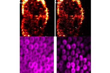
Wang and Yao subtracted the boundary area of the sample, leaving only the center — or what they call a photo imprint — now down to 80 nanometers wide, providing a very high, or super-resolution, picture. A smaller diameter of the center provides a better resolution in the image.
“In the end, we effectively shrink the detection spot to a smaller region,” Yao says. “Eighty nanometers allows us to see a lot of subcellular features, such as mitochondria or cell nuclei.”
After each area of the sample is scanned, the researchers create an image. With previous photoacoustic microscopy imaging, the microspheres on the image were blurry. However, with the new photo-imprint photoacoustic microscopy, the resulting image is clear and sharp.
“When we improve the resolution, we can see the cell structure with much more detail,” Yao says. “For biologists, these are much more informative images.”
Those working in imaging could apply this method to their own research, Yao says.
About The School of Engineering & Applied Science
The School of Engineering & Applied Science at Washington University in St. Louis focuses intellectual efforts through a new convergence paradigm and builds on strengths, particularly as applied to medicine and health, energy and environment, entrepreneurship and security. With 82 tenured/tenure-track and 40 additional full-time faculty, 1,300 undergraduate students, 700 graduate students and more than 23,000 alumni, we are working to leverage our partnerships with academic and industry partners — across disciplines and across the world — to contribute to solving the greatest global challenges of the 21st century.
Source: The School of Engineering & Applied Science
