New MRI Approach Could Detect Alzheimer's Disease Early
By Chuck Seegert, Ph.D.

Early detection of Alzheimer’s disease may now be possible based on research from a Northwestern University team. Their new magnetic resonance imaging (MRI) technique has been used to detect the earliest stages of the disease in a living animal, well before symptoms typically appear.
Alzheimer’s disease is an affliction that is prevalent in the aging population, and it takes a significant economic toll on society and a major emotional toll on the families of patients. Currently there are no methods for early detection of the disease. Conventional technology can identify amyloid plaques, an indicator that the disease has become advanced. Detection at this later stage allows intervention, but, when the disease is more advanced, treatment may only be marginally effective.
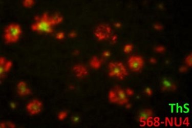
A new non-invasive MRI probe technology may overcome many of the drawbacks of existing diagnostics, according to a recent press release. Developed by engineers and scientists at Northwestern University, the new MRI probe is composed of nanoparticles attached to antibodies specific to amyloid beta oligomers. Amyloid beta oligomers are the toxic precursors to amyloid plaques and are now believed to attack synapses, destroying memory and eventually causing neurons to die. This biomarker may appear a decade or more before the appearance of amyloid plaques, which are caused by the accumulation and aggregation of the oligomers.
"We have a new brain imaging method that can detect the toxin that leads to Alzheimer's disease," said William L. Klein, a neuroscientist at Northwestern University, in the press release. "Using MRI, we can see the toxins attached to neurons in the brain. We expect to use this tool to detect this disease early and to help identify drugs that can effectively eliminate the toxin and improve health."
To test the new MRI probe, the team administered it to mice intranasally, according to a recent study published by the team in Nature Nanotechnology. Once placed onto the mucus membranes, the magnetic nanostructures traveled to the hippocampus, where they were bound to the amyloid beta oligomers by the antibodies. The MRI signal that the probes displayed was detected in diseased animals, but not in the control animals. When tested on samples of human brain tissue from Alzheimer’s patients, the probe was equally effective at highlighting amyloid beta oligomers.
While the treatment of Alzheimer’s is the focus of extraordinary research efforts, no effective treatment has been found, according to the press release. The new diagnostic probe, however, could enable these efforts by targeting changes in diseased tissues much earlier in the development of the illness.
"This MRI method could be used to determine how well a new drug is working," said Northwestern materials scientist Vinayak P. Dravid, according to the press release. "If a drug is effective, you would expect the amyloid beta signal to go down."
In addition to imaging the disease, observations during the study seem to indicate that the probe had potential for therapeutic intervention. Though it wasn’t a focus of the study, the researchers noticed the behavior of animals with Alzheimer’s seemed to improve after receiving only a single dose of the probe.
Detecting neurodegenerative diseases early on in their development may allow interventions at a time when they could have more impact on a patient’s health. Imaging advances for early detection are also being used to address other degenerative diseases like chronic traumatic encephalopathy.
Image Credit: ADAPTED FROM VIOLA ET AL., NATURE NANOTECHNOLOGY, 2014.
