Mini Gamma Ray Camera Offers Handheld Nuclear Medicine

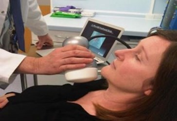
A team of researchers in the United Kingdom, working to translate space technology into biomedical applications, has designed a miniaturized gamma ray camera. They claim that, because the device is portable, it could be used to diagnose tumors and lymph nodes in intensive care units or operating rooms. Traditional gamma imaging has relied upon a much larger machine that scans the whole body and often occupies its own room.
John Lees, a research scientist working at the University of Leicester (UL), commented in a podcast that a great deal of technology from space could benefit the public. The handheld gamma ray camera, a collaboration between the University of Leicester and the University of Nottingham, is just one example. Lees believes that making the device portable will grant clinicians more diagnostic options and allow for earlier diagnosis of tumors than was previously possible.
“By significantly reducing the size of gamma cameras available we hope to provide far more flexibility for patients and clinicians — the camera doesn’t need a dedicated room and can be used by a patient’s bedside or even in the operating room,” said Sarah Bugby, a postgraduate researcher on Lees’ team, in a press release.
The camera combines optical and gamma imaging, and it offers a small scan field of view (SFOV). The camera works by superimposing a gamma-ray image on top of a visible image, which could allow a surgeon to more precisely locate and remove all cancerous lymph nodes before an operation concluded. This method, said researchers, “paves the way for less intrusive surgery and ensures all cancer cells are removed.”
Gamma probes currently used during surgical procedures, such as breast cancer sentinel node biopsies (SNB), rely on injected radioactive tracers and cannot provide imaging. This disadvantage, according to Lees, often leads to misdiagnosis.
In a study published last year in Physica Medica, researchers from Stanford University compared traditional gamma probes to the gamma ray camera during SNBs and discovered that the handheld camera located nodes that the probes had missed.
“Our system will improve surgical cancer treatments, reducing mortality and morbidity by enabling surgeons to increase lymph or tumor removal efficiency while minimizing damage to normal tissue,” said Lees in a press release.
The Space Research Center at the University of Leicester and Academic Medical Physics at the University of Nottingham have formed a new company called Gamma Technologies. Their goal is to design a line of portable devices that will bring nuclear medicine to the bedside, and the mini gamma ray camera is the first prototype to be clinically tested. They currently are investigating other applications for the device, including thyroid morphology, lymphatic drainage, and lacrimal drainage.
Alan Perkins, a professor of medicine at the University of Nottingham, commented, “This is an exciting project which is taking novel hybrid imaging technology into new clinical areas. This should expand the remit of nuclear medicine for the benefit of patients. Our preliminary clinical studies look very promising indeed.”
