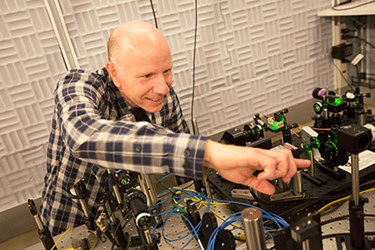
Harvard Scientists Create Nanoscale MRI
By Joel Lindsey

Researchers at Harvard have developed a magnetic resonance imaging (MRI) system that can generate nanoscale images, and could eventually reveal the atomic structures of individual molecules.
Details regarding the project have been released in an article published recently in the journal Nature Nanotechnology.
“What we’ve demonstrated in this new paper is the ability to get very high spatial resolution, and a fully operational MRI technology,” Amir Yacoby, Harvard professor of physics and of applied physics and lead researcher on the project, said in an article published by the Harvard Gazette. “This work is directed toward obtaining detailed information on molecular structure.”
“If we can image a single molecule and identify that there is a hydrogen atom here and a carbon atom there,” Yacoby added in the Gazette article, ”we can obtain information about the structure of many molecules that cannot be imaged by any other technique today.”
In order to develop the imaging device, Yacoby and his team essentially miniaturized a conventional MRI system.
“Functionally, it operates in the same way,” Yacoby said, “but in [miniaturizing it], we’ve had to change some of the components, and that has enabled us to achieve far greater resolution than conventional systems.”
The miniaturization was accomplished by using a tiny magnet — only 20 nanometers in diameter — capable of generating a magnetic field gradient 100,000 times larger than conventional X-ray systems, according to the Gazette article. The combination of its extremely small size and strong magnetic field enabled the device to be placed close to the object being imaged (within a few billionths of a meter), which allowed for spatial resolution of better than one nanometer.
In order to read the data produced from their tiny and powerful magnet, Yacoby’s team created qubits by embedding atomic-scale impurities called a nitrogen-vacancy (NV) centers in the “super-fine tips” of tiny milled devices. Qubits, or quantum bits, are the primary components of quantum computers.
Yacoby’s team found that when the device tip was scanned across the surface of a diamond crystal, the qubit interacted with the spinning motions of electrons near the crystal’s surface. These interactions could then be recorded and used to create an image of the electrons.
By bringing together the qubit sensor and newly designed nanoscale magnet, Yacoby and his team believe that it will be possible to generate detailed images of individual molecules’ constitutive components.
“Our current system is already capable of imaging individual electron spins with subnanometer resolution,” said Yacoby. “The goal, eventually, is to put a molecule in proximity to our NV center to try to see the components within that molecule, namely the nuclear spins of the individual atoms composing it.”
Image Credit: Kris Snibbe/Harvard Staff Photographer
