Can Retinal Imaging Predict Early-Stage Alzheimer's?
By Joel Lindsey

A group of researchers may have discovered a way to use a noninvasive retinal imaging device to reliably predict Alzheimer’s disease years before its actual onset.
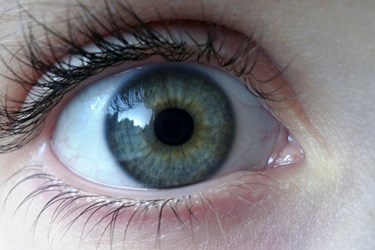
“The retina, unlike other structures of the eye, is part of the central nervous system, sharing many characteristics of the brain. A few years ago, we discovered at Cedars-Sinai that the plaques associated with Alzheimer’s disease occur not only in the brain but also in the retina,” Keith Black, professor and chair of Cedar-Sinai’s department of neurosurgery, said in a press release issued recently. “By ‘staining’ the plaque with curcumin, a component of the common spice turmeric, we could detect it in the retina even before it began to accumulate in the brain. The device we developed enables us to look through the eye — just as an ophthalmologist looks through the eye to diagnose retinal disease — and see these changes.”
Scientists have long linked buildups of beta-amyloid plaque in the brain with the development of Alzheimer’s, making the ability to observe the presence of this plaque a potentially powerful diagnostic tool. Traditional methods of diagnosing Alzheimer’s, however, can only detect beta-amyloid plaque after it has accumulated in the brain — usually when the disease has advanced to late stages.
Researchers at Australia’s Commonwealth Scientific and Industrial Research Organization (CSIRO) have used the device created by the Cedar-Sinai team to test the accuracy of using retinal imaging tests to predict Alzheimer’s.
In their tests, CSIRO scientists focused on trying to correlate retinal plaque buildups detected by optical imaging with brain plaque buildups detected by positron emission tomography (PET) scans. Finding such a correlation could help them gauge whether or not retinal imaging is a viable option for generating early predictions of whom may be at risk of developing Alzheimer’s.
They conducted tests on groups of patients already diagnosed with Alzheimer’s, patients with mild cognitive impairment, and a group of people currently without any evidence of brain abnormality.
“In preliminary results in 40 patients, the test could differentiate between Alzheimer’s disease and non-Alzheimer’s disease with 100 percent sensitivity and 80.6 percent specificity, meaning that all people with the disease tested positive and most of the people without the disease tested negative,” said Shaun Frost, a biomedical scientist and study manager at CSIRO. “The optical imaging exam appears to detect changes that occur 15-20 years before clinical diagnosis. It’s a practical exam that could allow testing of new therapies at an earlier stage, increasing our chances of altering the course of Alzheimer’s disease.”
In addition to possibly providing important early stage predictions, the tests could also represent a more convenient and accessible way to diagnose Alzheimer’s, according to the press release. Researchers pointed out that traditional Alzheimer’s tests rely on a PET scans, which require the use of radioactive tracers and the analysis of cerebrospinal fluids obtained through lumbar punctures. The new optical imaging approach is significantly simpler and noninvasive.
Details and results from the ongoing CSIRO study were described in a presentation at the Alzheimer’s Association International Conference 2014 in Copenhagen, Denmark.
