Are Piglet Brains The Key To Understanding Human Neurodevelopment?
By Chuck Seegert, Ph.D.

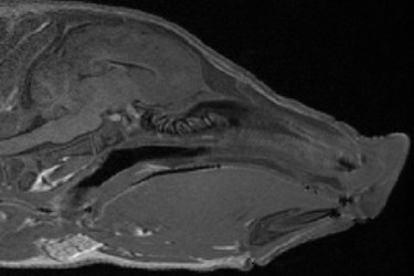
Using 15 piglet brains, researchers have developed a magnetic resonance imaging (MRI) model that is more detailed than ever before. The images were computationally averaged, and similarities to human brain development make it a viable reference for future studies.
Understanding normal neural development is a critical part of medicine, especially when attempting to understand what causes poor neural development. Finding effects on neural development caused by many environmental factors like illness and malnutrition are dependent on knowing what a normal level of development looks like. A control dataset is needed for comparisons like these.
MRI reference resources currently exist, but they are from adult pigs, which make them unsuitable for developmental studies, according to a recent press release from University of Illinois (UI). In many areas, the pig is thought to be an excellent translational animal model and is well representative of human development.
"The piglet brain is similar to the human brain in that it is gyrencephalic and experiences massive growth and development in the late prenatal and early postnatal periods,” said Rod Johnson, a UI professor of animal sciences, in the press release. “We are concerned that environmental insults such as infection or poor nutrition during these early periods may alter the trajectory of brain development."
The MRI atlas was created with images taken from 15, four-week old pigs. Volumes of 3D data were created from the images and, through advanced computational methods, the datasets were combined to create a final averaged brain model, according to a study published by the team in PLOS ONE. This well understood dataset will allow analysis tools like voxel-based morphology (VBM) to be implemented across many institutions, where before there was no reference that would allow them to be standardized.
"The benefit to using an averaged brain is that it will produce a template that is a better representation of the population,” said Matthew Conrad, a doctoral student in Johnson's lab, in the press release. “The more animals included the better."
Previous development studies used other, less effective methods besides VBM to look at iron deficiencies in brain development.
"For that we did MRI imaging and manual segmentation, and with manual segmentation you are looking at volume changes within very large areas of the brain, but with VBM we can pinpoint smaller changes within discrete brain areas," Conrad said in the press release. "We are now reanalyzing data from those piglets and replicating this study with new protocols, which will allow us to see changes that we didn't see before."
The MRI atlas is an open source reference material.
MRI studies are an invaluable tool in medical diagnostics and thus enjoy significant research support. Recently in an article published on Med Device Online, MRI methods with nanoscale resolution were reported.
Image Credit: Pig Imaging Group, University of Illinois
